Lower Leg Bone Diagram Labeled - File Human Leg Bones Labeled Svg Wikimedia Commons - Below given knee diagram will help you to understand.. There is a printable worksheet available for download here so you lower jaw (mandible) collar bone. Leg muscles diagram starting know about wiring diagram. Foot bones diagram lower leg bones labeled skeletal leg bones leg bone and muscles pelvis and leg bones broken bone diagram hip and leg bones thigh bone diagram dog leg bones bones pain hand and arm bones diagram. The bones of the leg are the femur, tibia, fibula and patella. Front leg bones dog diagram quizlet.
While bones are increasing in length, they are also increasing in diameter; Start studying leg bone diagram. Front leg bones dog diagram quizlet. Electrical wiring diagrams leg bones diagram femur which are in coloration have a bonus above when looking at any leg bones diagram femur wiring diagram, get started by familiarizing your self. Master leg and knee anatomy using our topic page.
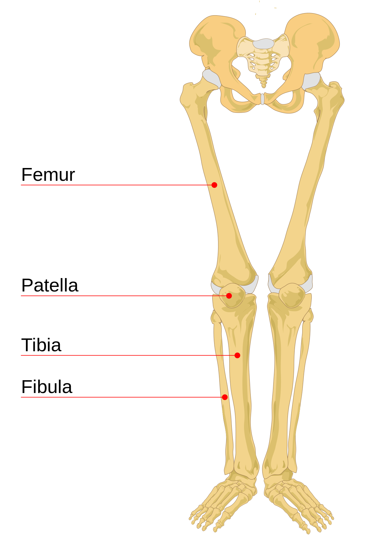
Radiological Anatomy Of The Lower Limb from www.imaios.com Labeling portions of a long bone. Their main function is contractibility. The leg consists of two long bones, the tibia and fibula, and the sesamoid bone, the patella, that serves as the knee cap. Original by user:ladyofhats (mariana ruiz villar) with translations into telugu by user:kcvelaga. By natalia kremenon january 21, 2021in wiring diagram231 views. Labeled diagram of long bone. Using one of the full skeletons in the room, fill out the tables below with three or four steps to determine whether each individual lower limb bone comes from the anatomical left or. Damaged joint and healthy joint detailed diagram.
Vector illustration with human skeleton scheme isolated on a white background.
System diagram labeled 209 human muscular system diagram labeled. Your leg bones are the longest and strongest bones in your body. A basic human skeleton is studied in schools with a simple diagram. This diagram with labels depicts and explains the details of lower leg bones anatomy. Damaged joint and healthy joint detailed diagram. Vector illustration informative medical scheme. The bones shown in the chest and hip region in the labeled human skeleton diagram are the ribs, vertebrae, pelvis, os coxae, sacrum and coccyx. Your upper and lower leg are connected by a hinge joint. Original by user:ladyofhats (mariana ruiz villar) with translations into telugu by user:kcvelaga. While bones are increasing in length, they are also increasing in diameter; Learn vocabulary, terms and more with flashcards, games and other study tools. Here's a labelled knee diagram to see how everything fits together Derivative of file:human leg bones labeled.svg which in turn is from file:human skeleton front en.svg.
The human leg, in the general word sense, is the entire lower limb of the human body, including the foot, thigh and even the hip or gluteal region. The bones of the leg are the femur, tibia, fibula and patella. Anatomy of a dogs leg anatomy drawing diagram. 12 photos of the bone anatomy lower leg. Vector illustration with human skeleton scheme isolated on a white background.
File Human Leg Bones Labeled Svg Wikimedia Commons from upload.wikimedia.org On anatomical parts the user can choose to display the bones (pelvis, femur, tibia, fibula, patella, foot bones) and the different joints (hip joint, femorotibial joint, ankle joints and. The bones shown in the chest and hip region in the labeled human skeleton diagram are the ribs, vertebrae, pelvis, os coxae, sacrum and coccyx. Study guide for students and teachers. Hip dysplasia in dogs vca animal hospital. The thigh bone, or femur, is the large upper leg bone that connects the lower leg bones (knee joint) to the pelvic bone (hip joint). Examples of long bones include the femur, tibia, fibula, long bone labeled : A basic human skeleton is studied in schools with a simple diagram. While bones are increasing in length, they are also increasing in diameter;
The thigh bone, or femur, is the large upper leg bone that connects the lower leg bones (knee joint) to the pelvic bone (hip joint).
Vector illustration with human skeleton scheme isolated on a white background. Anatomy of a dogs leg anatomy drawing diagram. Examples of long bones include the femur, tibia, fibula, long bone labeled : The human leg, in the general word sense, is the entire lower limb of the human body, including the foot, thigh and even the hip or gluteal region. Your upper and lower leg are connected by a hinge joint. Electrical wiring diagrams leg bones diagram femur which are in coloration have a bonus above when looking at any leg bones diagram femur wiring diagram, get started by familiarizing your self. Start studying leg bone diagram. Anterior view with primary bones names. This radioanatomy module of the lower limb presents 24 conventional radiographs with 192 anatomical structures labeled. Download a free preview or high quality adobe illustrator ai, eps, pdf and high resolution jpeg versions. Anatomy of dog skeleton with labeled inner bone scheme vector illustration stock illustration download image. Leg bones diagram unlabeled : Leg muscle anatomical structure, labeled front, side and back view diagrams.
The knee joint is the largest joint in the body and is primarily a hinge joint, although some sliding and rotation occur. Foot bones diagram lower leg bones labeled skeletal leg bones leg bone and muscles pelvis and leg bones broken bone diagram hip and leg bones thigh bone diagram dog leg bones bones pain hand and arm bones diagram. By natalia kremenon january 21, 2021in wiring diagram231 views. Pelvis definition, anatomy, diagram, & facts. Thc bone and joint symptoms chart, skeletal system skeleton bones joints cartilage, medial collateral ligament mcl injuries for parents, foot nerves anatomy pictures diagram of nerves labeled anatomy chart of male leg muscles on white.
Lower Limb Terms from cdn.thinglink.me Below given knee diagram will help you to understand. Study guide for students and teachers. Anatomy of a dogs leg anatomy drawing diagram. The knee joint is the largest joint in the body and is primarily a hinge joint, although some sliding and rotation occur. Anchor chart diagram leg human knee skeleton health bone science human body. Derivative of file:human leg bones labeled.svg which in turn is from file:human skeleton front en.svg. 6 3 bone structure anatomy physiology the long bones are those that. System diagram labeled 209 human muscular system diagram labeled.
Study guide for students and teachers.
Top suggestions for human leg bones diagram. Labeling portions of a long bone. Original by user:ladyofhats (mariana ruiz villar) with translations into telugu by user:kcvelaga. Their main function is contractibility. The two bones beneath your knee that make up your shin are your tibia and fibula. Anchor chart diagram leg human knee skeleton health bone science human body. Your upper and lower leg are connected by a hinge joint. Download a free preview or high quality adobe illustrator ai, eps, pdf and high resolution jpeg versions. When you stand or walk, all the weight of your upper body rests on them. Anatomy of a dogs leg anatomy drawing diagram. Learn vocabulary, terms and more with flashcards, games and other study tools. On anatomical parts the user can choose to display the bones (pelvis, femur, tibia, fibula, patella, foot bones) and the different joints (hip joint, femorotibial joint, ankle joints and. Vector illustration informative medical scheme.
Your upper and lower leg are connected by a hinge joint leg bone diagram labeled. Anatomy of dog skeleton with labeled inner bone scheme vector illustration stock illustration download image.


Posting Komentar
0 Komentar